Showing posts with label eye. Show all posts

Tuesday, October 6, 2015

New gene therapy for vision loss from a mitochondrial disease

NIH-funded study shows success in targeting mitochondrial DNA in mice
Researchers funded by the National Institutes of Health have developed a novel mouse model for the vision disorder Leber hereditary optic neuropathy (LHON), and found that they can use gene therapy to improve visual function in the mice. LHON is one of many diseases tied to gene mutations that damage the tiny energy factories that power our cells, called mitochondria.
Image of successful delivery of mtDNA to mouse retina
Dr. John Guy and colleagues added a homing signal to a virus in order to deliver the ND4 gene into mitochondria. A marker for the gene is shown in red and the ND4 protein is shown in green, inside retinal ganglion cells in the mouse eye. The nuclei of retinal ganglion cells are shown in blue. Credit: Dr. Hong Yu, Bascom Palmer.
“This study marks an important contribution to research on LHON, and in efforts toward an effective therapy. But the implications are even broader, because the approaches that the investigators used could aid therapy development for a vast array of other mitochondrial diseases,” said Maryann Redford, D.D.S, M.P.H., a program director in Collaborative Clinical Research at NIH’s National Eye Institute, which helped fund the study.
Mitochondria are as complex as any modern manufacturing facility, with specialized machinery for converting nutrients and oxygen into cellular energy. They even have their own DNA, and it is mutations within this mitochondrial DNA (mtDNA) that lead to LHON, as well as a host of other diseases. But the unique nature of mtDNA has presented challenges for developing and testing potential therapies for such diseases.
Until now, “there was no efficient way to get DNA into mitochondria,” said John Guy, M.D., professor of ophthalmology and director of the ocular gene therapy laboratory at the Bascom Palmer Eye Institute, University of Miami Miller School of Medicine. Dr. Guy’s laboratory is among the first to develop an approach that can target mtDNA in living mice and people.
Their success in creating a mouse model of LHON and using it to test an investigational gene therapy is described today in the Proceedings of the National Academy of Sciences.
The global impact of LHON is unknown. In England, the estimated prevalence is about 1 in 30,000. Early symptoms include blurry vison and usually appear during the teens or early twenties. Eyesight tends to worsen over time, eventually leading to a severe loss of sharpness (acuity) and color vision. These problems are caused by a loss of retinal ganglion cells — the cells that carry visual signals from the retina through the optic nerve and into the brain.
The most common mutation behind LHON impairs a mitochondrial gene called ND4. Dr. Guy began to research a possible gene therapy approach for delivering a substitute copy of the gene into mitochondria about 15 years ago. In most studies and applications of gene therapy, viruses have become the preferred vessel for delivering genes into cells. But viruses evolved to invade the body’s cells and penetrate the nucleus, which contains the bulk of our DNA, comprising about 20,000 genes. Most viruses are poor at penetrating mitochondria.
To fix that, Dr. Guy and his team took advantage of the fact that mitochondria import cellular proteins that they cannot make themselves. By attaching a bit of one such protein to the outer shell of a virus — called an adeno-associated virus — he effectively gave the virus a homing signal and entry code into mitochondria.
This modified virus has been the key to creating a mouse that replicates LHON and to an investigational gene therapy for LHON that is currently in clinical trials.
To create a mouse model for LHON, the researchers loaded the virus with a defective copy of the ND4 gene carrying the same mutation that causes about 70 percent of LHON cases. They also included DNA coding for a red fluorescent protein, as a visible marker for the virus and its payload. Then they injected the virus into fertilized mouse egg cells, and grew the cells to maturity.
After breeding the mice through several generations, the researchers had their mouse model. The presence of the virally encoded ND4 mutation in the eye was confirmed by essentially doing an eye exam to look for the red fluorescent marker. Over time, the mice showed a loss of retinal ganglion cells, atrophy (shrinkage) of the optic nerve, and a decline in visual responses, as seen in a type of electrical recording from the retina known as an electroretinogram.
To develop a gene therapy for LHON, the team packaged the normal human ND4 gene into the same stealthy virus. This combination, when injected into the eye, led to improved visual function in the LHON mouse model. When injected into normal mice, the virus carrying ND4 did not cause any adverse effects on vision.
Prior to development of the new mouse model, Dr. Guy’s lab had shown that they could produce temporary signs of LHON in mice. They were able to prevent development of LHON in the mice, but not reverse it. “Now we’ve shown that we can improve visual function after it’s been lost,” he said.
The mouse research is helping inform an ongoing NEI-supported clinical trial, which is led by Dr. Guy and is testing the safety of the same gene therapy approach (without the red fluorescent protein) in people with LHON. The trial is recruiting LHON patients who fit into three categories — those with chronic vision loss in both eyes, with recent-onset vision loss in both eyes, or with recent-onset vision loss in one eye but no signs of abnormal vision in the other eye. For more information, visit http://www.clinicaltrials.gov and search for the trial identifier NCT02161380.

Tuesday, September 1, 2015

How the eye sees small moving objects

NIH-funded study reveals how motion-sensing cells in mice link to other cells in the eye
When we move our head, the whole visual world moves across our eyes. Yet we can still make out a bee buzzing by or a hawk flying overhead, thanks to unique cells in the eye called object motion sensors. A new study on mice helps explain how these cells do their job, and may bring scientists closer to understanding how complex circuits are formed throughout the nervous system. The study was funded by the National Institutes of Health, and was published online in Nature.
“Understanding how neurons are wired together to form circuits in the eye is fundamental for advancing potential new therapies for blinding eye diseases,” said Paul A. Sieving, M.D., Ph.D., director of NIH’s National Eye Institute (NEI). “Research aimed at regenerating photoreceptors, for example, is enriched by efforts to understand the circuitry in the eye.”
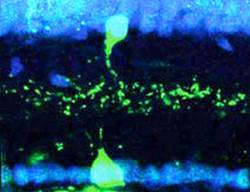
Object motion and amacrine cells in retina
A VG3 amacrine cell (top, in green) is shown superimposed with an object motion detector cell (bottom, green). Each of the neurons is expressing sidekick-2, an adhesion molecule that allows the two neurons to find each other and connect.
Object motion sensors are one of about 30 different types of retinal ganglion cells (RGCs) in the retina, the light-sensitive tissue at the back of the eye. These cells are unique because they fire only when the timing of a small object’s movement differs from that of the background; they are silent when the object and the background move in sync. Researchers believe this is critical to our ability to see small objects moving against a backdrop of complex motion.
The cells in the retina are wired up like an electrical circuit. Vision begins with photoreceptors, cells that detect light entering the eye and convert it into electrical signals. Middleman neurons, called interneurons, shuttle signals from photoreceptors to the RGCs. And each RGC sends the output visual information deeper into the brain for processing. This all takes place within fractions of a second, so the scientists were surprised to discover that before it reaches object motion sensors, visual information about object motion takes a detour through a unique type of interneuron. Their results represent an ongoing effort by scientists to map out complex circuits of the nervous system.
“Getting these connections precisely correct is incredibly important, as each specific feature of vision, such as seeing a particular direction of motion or a color, relies on it,” said the study’s lead investigator, Arjun Krishnaswamy, Ph.D., of Harvard University’s Center for Brain Science, Cambridge, Massachusetts. “It’s also incredibly complex. Within the retina, all these different types of RGCs and interneurons intermingle as they develop. There have to be remarkable ways to sort them out so they connect up properly.”
Using a genetically engineered mouse line, the researchers recorded the activity of object motion sensors and found that the cells form synapses (or connections) with interneurons called VG3 amacrine cells. What’s interesting about this connection is that most retinal circuits tend to follow a more direct, and therefore faster, route. RCGs typically are two synapses away from a photoreceptor, but with the addition of VG3 amacrine cells to the circuit, object motion sensors appear to be three synapses away, slowing visual information delivered to the cells.
To test this idea, the scientists flashed light on the retinas of the mice and found that on average the object motion sensors responded later than other types of retinal ganglion cells. They also selectively activated the sensors by projecting light patterns onto the retinas that mimicked the movement of small objects against a desynchronized background. Mice with genetically eliminated VG3 amacrine cells did not show these responses.
The researchers theorize that the longer pathway contributes to an essential delay, ensuring that information from the central field of view and from the periphery arrive at the object motion sensor at the same time. This in turn allows the object motion sensors to accurately assess the difference between the motion of a hawk and the slow-moving clouds above it, or the flight of a baseball and the undulating crowd in the stadium.
Dr. Krishnaswamy and his colleagues then investigated how the circuit develops. They found that object motion sensors and VG3 amacrine cells each make a protein called sidekick-2, specifically where they contact each other. Sidekick-2 is an adhesion molecule that allows the two cell types to find each other and stick together so they can communicate across a synapse.
Mice genetically engineered to block sidekick-2 production lacked synapses connecting VG3 cells to object motion sensors. Moreover, electrical recordings showed that sensors in these mice did not distinguish the motion of a small object from background motion.
For Dr. Krishnaswamy, the next steps are to investigate the role of sidekick-2 in brain development, first in mice and eventually in humans.
“Neurons in the brain work just like neurons in the retina, and scientists are beginning to understand how they make precise connections to form circuits that control thoughts, senses and emotions,” said Edmund Talley, Ph.D., program director at the National Institute of Neurological Disorders and Stroke (NINDS), part of the NIH. “This pioneering work demonstrates the molecular specificity behind these connections.”