Showing posts with label MRI. Show all posts

Wednesday, October 21, 2015

Cardiovascular Research:Botox may prevent irregular heartbeat after bypass surgery ♦ MRI shows heart ages differently in women than in men

Pacemakers identify atrial fibrillation and enable initiation of stroke prevention Pacemakers identify atrial fibrillation and enable initiation of anticoagulation to prevent strokes.
Boosting levels of a key growth factor may help prevent cardiovascular disease New research indicates that low levels of a growth factor called stem cell factor (SCF)—which is thought to be important for blood vessel repair—are linked with an increased risk of cardiovascular disease. In a study of 384 patients with a first coronary event and 409 event-free individuals, SCF measurements had a predictive value that was in the same range as that of established cardiovascular risk factors such as LDL and HDL cholesterol. Moreover, smokers and diabetics had reduced levels of SCF in the circulation, and low blood levels of the growth factor were associated with more severe disease among patients with atherosclerosis.
Botox may prevent irregular heartbeat after bypass surgery Botox injections into fat surrounding the heart reduced the chances of developing an irregular heart rhythm -- the most common complication of bypass surgery.
MRI shows heart ages differently in women than in men The main pumping chamber of the heart ages differently in men and women, according to a MRI study. Researchers said the findings may support different treatment approaches for men and women with heart disease.

Wednesday, September 30, 2015

Women's Health: Maintaining healthy DNA delays menopause ♦ MRI technique could reduce need for breast biopsies ♦ Microchip used to visualize human breast cancer proteins

Maintaining healthy DNA delays menopause An international study of nearly 70,000 women has identified more than 40 regions of the human genome involved in governing at what age a woman goes through menopause. The study found that two-thirds of those regions contain genes that act to keep DNA healthy. It also found the first genetic evidence of a link between the timing of menopause and breast cancer, corroborating previous conclusions from observational evidence.
Scientists use microchip approach to visualize human breast cancer proteins Scientists present a new molecular toolkit to investigate protein assemblies natively formed in the context of human disease. BRCA1 gene regulatory complexes from cancer cells were visualized for the first time.
How more women with earlier Cesarean sections can give birth vaginally next time In many countries, caesarean section is routinely used if the woman previously gave birth by caesarean section. Doctors and midwives in countries with a high rate of vaginal births after caesarean sections have for the first time been asked in a study to give their views on how to increase the percentage of vaginal births.
Biomarkers in maternal blood can identify pregnant women with lupus at low risk for adverse outcomes Pregnant women with systemic lupus erythematosus, are at higher risk for adverse pregnancy outcomes, including preeclampsia, placental insufficiency, fetal death, miscarriages, and other complications. A consortium of top researchers reports that monitoring specific angiogenic biomarkers in maternal blood during early pregnancy can successfully predict patients who will likely have normal pregnancies.This will enable physicians to identify, counsel, and manage high risk patients at an early stage of pregnancy.

MRI technique could reduce need for breast biopsies A magnetic resonance breast imaging technique that uses no ionizing radiation or contrast agent could reduce unnecessary biopsies by providing additional information about suspicious findings on X-ray screening mammography

Sunday, June 7, 2015

Brain Research: Evidence of schizophrenia's causes ♦ Hippo-campus: In search of memory storage ♦ Long-term memory formation

MRI technology reveals deep brain pathways in unprecedented detail A 3-D map of the human brain stem has been produced at an unprecedented level of detail using MRI technology. In a new study, the researchers unveil an ultra high-resolution brain stem model that could better guide brain surgeons treating conditions such as tremors and Parkinson's disease with deep brain stimulation
Scientists produce strongest evidence yet of schizophrenia's causes The strongest evidence yet of what causes schizophrenia has been provided by an international group of scientists. The work strongly suggests that disruption of a delicate chemical balance in the brain is heavily implicated in the disorder.
Hippocampus: In search of memory storage The hippocampus plays a crucial role in memory formation. However, it is not yet fully understood in what way that brain structure's individual regions are involved in the formation of memories. Neuroscientists have recreated this process with the aid of computer simulations. Their findings challenge the model of memory forming in the hippocampus
Autologous stem cell therapy helpful in traumatic brain injury The use of cell therapy after traumatic brain injury in children can reduce the amount of therapeutic interventions needed to treat the patient, as well as the amount of time the child spends in neurointensive care, according to research.
Long-term memory formation Neuroscientists has determined how a pair of growth factor molecules contributes to long-term memory formation The researchers examined GFs in Aplysia californica, the California sea slug. Aplysia is a model organism that is quite powerful for this type of research because its neurons are 10 to 50 times larger than those of higher organisms, such as vertebrates, and it possesses a relatively small network of neurons -- characteristics that readily allow for the examination of molecular signaling during memory formation.

Thursday, May 28, 2015

Health News: 34 Sickened in Canadian salmonella outbreak ♦ Job-sharing with nursing robots ♦ New technique speeds nano MRI imaging

34 Sickened in Canadian salmonella outbreak linked to chicks from Alberta hatchery An investigation is underway in Canada to figure out the source of a Salmonella Enteritidis outbreak linked to live chicks from an Alberta hatchery. The outbreak has so far sickened 34 people in three provinces
Job-sharing with nursing robots Given the threat of a massive earthquake striking Japan, researchers have organized a cooperative project team to develop a new robot. They set about determining the factors that are most important to hospital patients in modern society, after which they set about developing the world's first medical round robot capable of job-sharing, making more time for face-to-face nursing care.
New urine test could reduce need for blood samples If you've been to the doctor, you probably know what to do when you're handed a plastic cup and shown to the bathroom. Most patients hand over the sample and give little thought to what happens when it's shipped to the lab for analysis. Researchers have developed a new testing method that they believe will reduce costs, get faster results and lower the volume of urine needed for a sample.
New technique speeds nano MRI imaging Nano MRI is a scanning technique that produces nondestructive, high-resolution 3-D images of nanoscale objects, and it promises to become a powerful tool for researchers and companies exploring the shape and function of biological materials such as viruses and cells in much the same way as clinical MRI today enables investigation of whole tissues in the human body.
Subclinical hyperthyroidism associated with an increased risk of hip and other fractures In an analysis that included more than 70,000 participants from 13 studies, subclinical hyperthyroidism was associated with an increased risk for hip and other fractures including spine. Subclinical hyperthyroidism is a low serum thyroid-stimulating hormone concentration in a person without clinical symptoms and normal thyroid hormone concentrations on blood tests.

Friday, May 15, 2015

Cancer Research:Obesity increases risk of developing cancer ♦ New targets for cancer treatments ♦ Taking immortality away from cancer

Potential new targets for cancer treatments identified Key steps in cell division have now been revealed by new research, possibly leading to treatments aimed at inhibiting cancer cell division,
Researchers take step toward bringing precision medicine to all cancer patients Researchers have developed and tested a new tool that searches for the most common genetic anomalies seen in cancer. The assay demonstrates the ability to make gene sequencing easier over a large volume of samples.
MRI shows potential to improve breast cancer risk prediction Magnetic resonance imaging (MRI) provides important information about a woman's future risk of developing breast cancer. The findings support an expanded role for MRI in more personalized approaches to breast cancer screening and prevention.
Obesity increases risk of developing cancer Cancer is more likely to develop in people who are very overweight (obese), because surplus body fat interferes with various hormone cycles and with glucose and fat metabolism. A metabolic expert draws attention to the fact that more and more people are suffering from obesity, spurring several studies to investigate the links between obesity, metabolic disorders and cancer
Taking immortality away from cancer Researchers report that they have blocked the shelterin protein TRF1 in cancer cells, disrupting telomere protection and preventing immortal proliferation of cancer cells. Their work shows for the first time that telomeres -- the structures protecting the ends of the chromosomes -- may represent an effective anti-cancer target: by blocking the TRF1 gene, which is essential for the telomeres, they have shown dramatic improvements in mice with lung cancer.

Wednesday, May 13, 2015

A SMARTer approach to stroke care

NIH study uses MRI scans to screen stroke patients for acute treatment within the first critical 60 minutes
Time is critical when it comes to stroke, and early treatment is associated with better outcomes. According to the Screening with MRI for Accurate and Rapid stroke Treatment (SMART) study, small changes in quality improvement procedures enabled clinicians to use MRI scans to diagnose stroke patients before giving acute treatment, within 60 minutes of hospital arrival. MRI scans provide detailed images but take longer to complete than CT scans, which are commonly used in most centers. The findings, published in Neurology, were supported in part by the National Institutes of Health’s National Institute of Neurological Disorders and Stroke (NINDS).
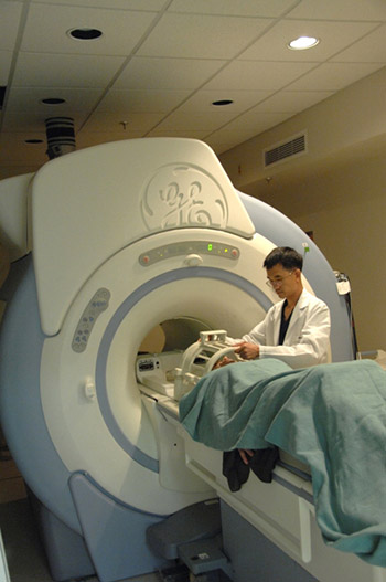
Image of a doctor and patient using an MRI.
New findings suggest that MRI machines (such as the one pictured above) may help quickly screen stroke patients for acute treatment. Image courtesy of the National Institutes of Health.
“By making small changes to our processes, we were able to scan suspected stroke patients with MRI and appropriately treat patients within a goal time of 60 minutes. This is an important finding for hospitals, healthcare providers and the public,” said Amie Hsia, M.D., medical director of the Comprehensive Stroke Center at MedStar Washington Hospital Center, Washington D.C., and senior author of the study. “Not only does MRI provide more precise and complete information than the traditionally used CT scan, now we’ve also demonstrated that it is feasible to use from a time perspective.”
National guidelines suggest that stroke patients should receive treatment within 60 minutes of arriving at the hospital. The majority of hospitals rely on rapid CT scans to determine if an individual is eligible for intravenous tPA, the only FDA-approved treatment for ischemic strokes, those caused by blood clots in the brain. If a CT scan shows the patient is having a bleeding, or hemorrhagic, type of stroke, tPA cannot be used as treatment. For many years CT scans were the only imaging tool available in most hospitals, but now MRIs are becoming more widely available.
Clinicians at MedStar Washington Hospital Center and Suburban Hospital, Bethesda, Maryland, routinely work with physician-scientists from NIH and have access to their cutting-edge medical protocols and technologies. The two hospitals use MRI instead of CT scans to screen stroke patients. Although MRI scans can take up to 15 minutes longer than CT scans, they provide clinicians with more detailed information about what is happening in a patient’s brain. Using MRI, clinicians can see early changes taking place during the stroke. In this way, they can see what tissue is at risk and identify blocked blood vessels or subtle bleeding that cannot be picked up by CT.
To reduce the door to treatment time, multidisciplinary teams at both hospitals carefully examined the existing processes to identify time-consuming bottlenecks or duplicative methods. By using “lean process interventions,” they found a number of steps that could be eliminated or changed. For example, at MedStar Washington Hospital Center, a lengthy MRI screening form was simplified to three questions; at Suburban Hospital, tPA was put into the medication cart in the MRI suite so that it could be given immediately to patients after scanning instead of returning them to the Emergency Department for treatment.
Once the changes were implemented, Dr. Hsia’s team examined whether they had an impact on treatment times for patients. The results indicated that door to treatment time was reduced from 93 to 55 minutes, a difference of 40 percent. Over a two year period, the percentage of patients treated within 60 minutes increased from 13 to 61.5 percent. Further analysis revealed that these changes were due to faster MRI start times.

Wednesday, May 6, 2015

Health Research: New strategies to control diabetes ♦ Who benefits from a catheter -- and who doesn't? ♦ Chickenpox virus can cause a stroke in an HIV patien


Chickenpox virus can cause a stroke in an HIV patient Varicella-zoster virus causes chickenpox in children and shingles in older adults. The virus typically remains dormant in patients with healthy immune systems, but can reactivate. Patients infected with human immunodeficiency virus (HIV) can, in rare cases, experience bleeding on the brain that causes a type of stroke called intracerebral hemorrhage.
Who benefits from a catheter -- and who doesn't? New guide aims to protect patients What's the only thing worse than having a urinary catheter when you're in the hospital? Having one and getting a urinary tract infection -- or worse -- as a result. A new detailed guide gives doctors and nurses information to help decide which hospital patients may benefit from a urinary catheter.
Redesigned systems may increase access to MRI for patients with implanted medical devices New technology may extend the benefits of magnetic resonance imaging to many patients whose access to MRI is currently limited. A redesign of the wire at the core of the leads that carry signals between implanted medical devices and their target structures significantly reduces the generation of heat that occurs when standard wires are exposed.
Molecular link between high glucose, metabolic disease may offer new strategies to control diabetes Scientists say they've discovered a cause-and-effect link between chronic high blood sugar and disruption of mitochondria, the powerhouses that create the metabolic energy that runs living cells. The discovery sheds light on a long-hidden connection and could lead to new ways of preventing and treating diabetes.
Online training can teach psychotherapists evidence-based treatments, Psychotherapy treatments can lag years behind what research has shown to be effective because there simply are not enough clinicians trained in new methods. A new study proposes one solution to the problem. The findings show that employing online training programs to teach psychotherapists how to use newer evidence-based treatments.

Tuesday, March 24, 2015

Health News: Opossum-based antidote could save thousands of lives ♦ Fruit likely caused listeria ♦ Vitamin D helps immune cells prevent atherosclerosis, diabetes + more

CDC:Recalled fruit likely caused listeria cases last summer  This past summer, consumers were confronted with a series of recalls of peaches, nectarines, plums, pluots and other stone fruits potentially contaminated with Listeria monocytogenes. The fruit, sourced from Wawona Packing Co., was recalled from major retailers such as ALDI, Trader Joe’s, Costco, Kroger and Walmart.. Continue Reading
Being near greened vacant lots lowers heart rates  Greening vacant lots may be associated with biologic reductions in stress, according to a new study. Residents who walked near newly greened vacant lots had significantly lower heart rates compared to walking near a blighted, or neglect Continue Reading
Vitamin D helps immune cells prevent atherosclerosis, diabetes Altered signaling through the vitamin D receptor on certain immune cells may play a role in causing the chronic inflammation that leads to the combination of type 2 diabetes and heart disease. These are the most common cause of illness and death Continue Reading
Opossum-based antidote to venom from snake bites could save thousands of lives Scientists have turned to the opossum to develop a promising new and inexpensive antidote for venomous snake bites. They predict it could save thousands of lives worldwide without the side effects of current treatments. Continue Reading
Total body iron balance: Liver MRI better than biopsy  Investigators have demonstrated that MR imaging of the liver is more accurate than liver biopsy in determining total body iron balance in patients with sickle cell disease and other disorders requiring blood transfusion therapy. This discovery follows the researchers earlier work in pioneering techniques to use MRI to noninvasively measure liver iron. Continue Reading

Friday, September 5, 2014

FDA Explores New Uses for MRI Scans


Some say a picture is worth a thousand words, but when that picture is used to study a brain, its worth is immeasurable.
Creating such images is the task of Sergei Liachenko, M.D., Ph.D., director of the bio-imaging program at the Food and Drug Administration’s National Center for Toxicological Research. Working in a small lab on the NCTR campus south of Little Rock, Ark., Liachenko and his team, in collaboration with the Center for Drug Evaluation and Research, are studying the effects of various chemicals on a live rat’s brain. Their goal is to prove that studying a live animal with a research-grade magnetic resonance imaging device can be at least as accurate, if not more so, as studies performed under current research procedures.
“We are trying to demonstrate that imaging can be used for testing physical damage to the brain, known as neurotoxicity,” Liachenko says, “and when it comes to studying neurotoxicity, the more detailed a picture we can take, the better.”
That’s where MRI comes in. It provides a three-dimensional and very sophisticated view of the brain.
How Does MRI Work and What are Its Benefits?
MRI machines use very powerful magnets to bring the water in the body’s cells to a “resonant” state. The machine then sends a resonating radio frequency, which travels through the body and is reflected back. The machine uses the information from this reflection to form a three-dimensional image of the area being scanned. MRI is widely used in hospitals to help identify diseases.
Currently, most scientists look for the damaged brain tissue by sampling multiple pathology slices of the brain of a deceased animal. But Liachenko and his CDER colleague Joseph Hanig are concerned this method could miss some damage because the entire brain is not tested. By contrast, MRI can picture the whole brain in contiguous slices, allowing every part to be examined for changes or damage.
“This assessment allows the entire brain to be scanned and produced in a 3-D representation, so we can view the tissues in different projections, and we don’t need to worry about missing potential damage because it’s in an area that was not examined,” says Liachenko.
Another benefit to using an MRI device is the ability to test a single rodent efficiently, repeatedly and safely. “It’s just a magnetic field, and we’re using what’s considered the safest imaging modality. That’s why we can put the same animal in the machine many times,” Liachenko says.
Studying the same animal over a long period of time, Hanig adds, “gives us more data that’s arguably more accurate than data from multiple subjects.” In addition, these MRI assessments are similar to those made in humans and therefore they could be connected with clinical research.
Yet another important advantage of using the MRI is that the imaging takes hours to complete and analyze, while conventional histological analysis procedures take weeks. This change in time frame, along with fewer subjects being used, could make imaging a more cost effective option.
Imaging at FDA
MRI is used all over the globe to investigate live animals, but what is unique to NCTR, in collaboration with CDER, is its study to develop MRI biomarkers of neurotoxicity. By using a set of compounds that cause known damage to the brain, the researchers can check the efficiency of the MRI scans compared to the conventional methods by making sure the MRI machine finds the same problems, and perhaps even more.
“Should the biomarkers prove to be reliable and the qualification is met, it will send a huge message to the pharmaceutical community that this method is good for assessing for potential brain damage,” Liachenko says. “In our view, this MRI research will create the opportunity to provide much safer drugs to people, and that’s the reason for our work.”